The '5 Main Muscles of Movement' Made Easy.
post by leggi · 2019-12-07T09:17:50.359Z · LW · GW · 0 commentsContents
  1. Rectus femoris
    The rectus femoris muscles align the hip and knee joints.
  2. Gluteus maximus.
    The gluteus maximus works in tandem with the rectus femoris, stabilising the legs to the torso (hip, pelvis to femur) and leading the legs through a full range of natural movement - when connected to Base-Line support.
  3. Pelvic floor. BASE
    The pelvic floor muscles.
    The base of the body.
  4. Rectus abdominis. LINE
    Feel your rectus abdominis muscles:
    The rectus abdominis muscles - our core pillar of strength from where all movement should originate.
      Breathe with your Base-Line.
      Think stronger and longer with every breathe in.
  5. Trapezius
        Movement of the upper body should begin from the lower trapezius.
    The trapezius muscles - guiding and supporting the head and arms through a full range of movement and aligning the upper body.
    BODY ALIGNMENT with the main muscles of movement:
    Time and Effort Required.
None
No comments
A post to help find the 5 main muscles (from my Base-Line Theory of Health and Movement) on your own body.
Anatomy is wordy. It's easy to get lost, but knowing the details isn't important:
- Study the pictures. See.
- Palpate your body. Feel.
Keep thinking about these 5 (paired - left and right) 'main muscles of moment' and how you use them as you move through your daily life.
Get to know how your body feels.
Become more aware of your posture [LW · GW] (the position of all of your body, all of the time).
Keep in mind images and diagrams aren't what the tissues of the body are really like. We are made up of a web of connective tissues, in many forms, within which the cells our organs and muscles are contained. Structures are not isolated parts, our body blends from one named structure to another.
1. Rectus femoris
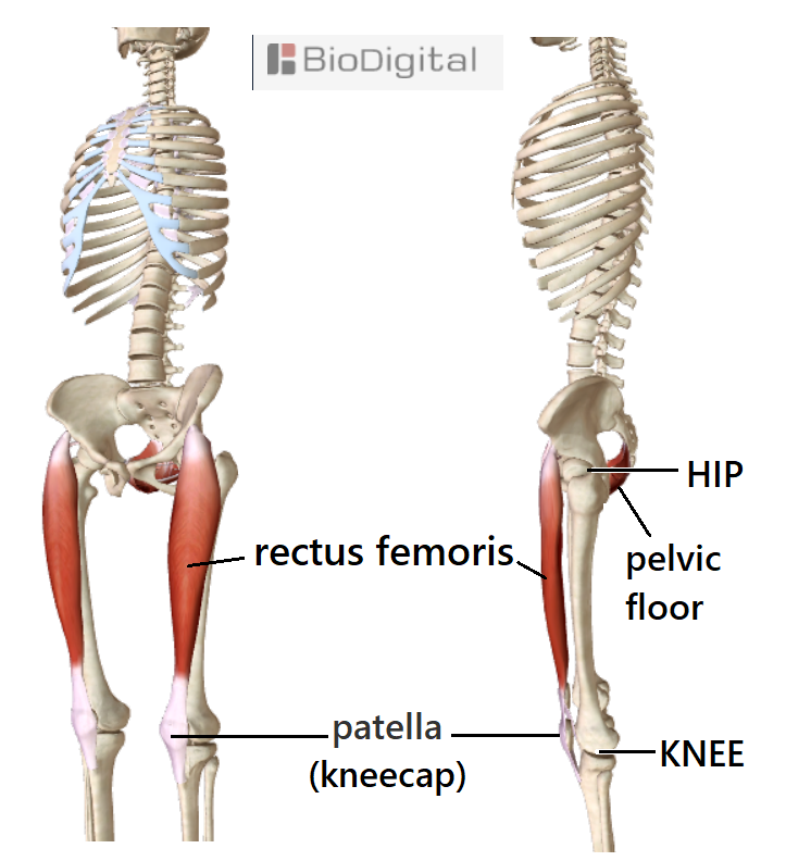
Below the knee, feel for the lump (tibial tuberosity) at the front of your shin bone (tibia). Run your hands up over your kneecaps and front of your thighs to just below the sticking-out bone at the front of your pelvis (hip bone). This is the full extent of the rectus femoris muscle.
- Aim for the whole muscle to be active.
- A strong, straight pole at the front of each thigh from hip to shin.
- Think of pulling your kneecaps up to activate the muscle + a downward extension from your hip bone.
From shin - a ligament that contains the kneecap turning into a layer of connective tissue at the back of the muscle.
From hip - short ropes of tendon from the hip bones that turn into a layer of connective tissue down the front of the rectus femoris muscles.
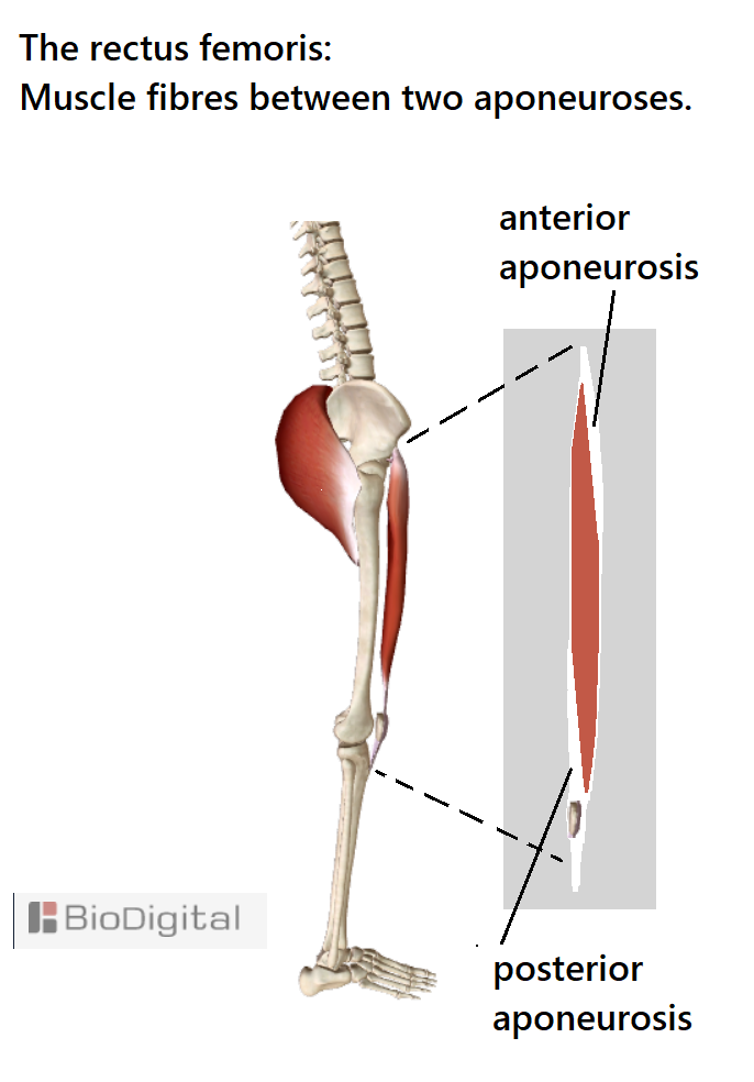
The rectus femoris is part of the quadriceps muscle group but it is the only one of the 4 muscles to attach to the pelvis - the other 3 attach to the top of the femur and thus do not cross the hip joint.
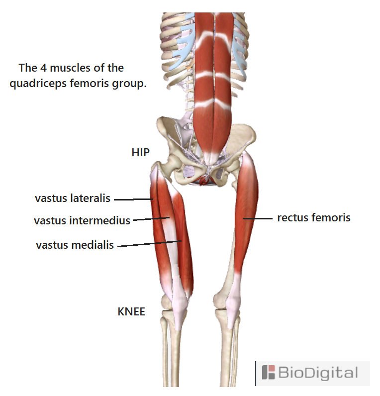
The rectus femoris muscles align the hip and knee joints.

2. Gluteus maximus.
The largest skeletal muscles of the body (covering a lot of complicated anatomy prone to pain/injury).

Look at the position of the hip joint in relation to the gluteus maximus. The "ball and socket" joint sits centrally in front of the gluteus maximus.
The gluteus maximus has a linear attachment to the top third of the back of the femur (thigh bone).
Hands on buttocks - feel for the muscles contracting. "Buns of steel", extending to the femur.
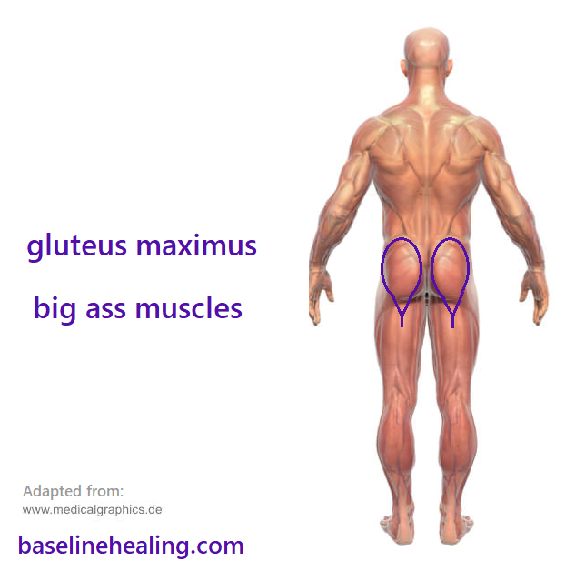
The gluteus maximus works in tandem with the rectus femoris, stabilising the legs to the torso (hip, pelvis to femur) and leading the legs through a full range of natural movement - when connected to Base-Line support.
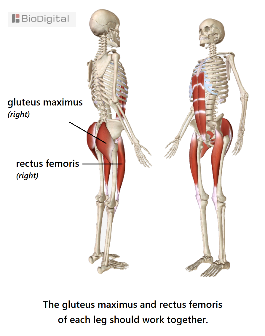
Now the key muscles to connecting mind to muscles, body to brain ... The body's 'core pillar of strength' and key to feeling your state of physical alignment and balance: Your Body's 'Base-Line':
3. Pelvic floor. BASE
The pelvic floor - a basket of muscles within the bones of the pelvis.


The pelvic floor consists of several small muscles spanning the pelvic canal. Left and right sides a mirror image.
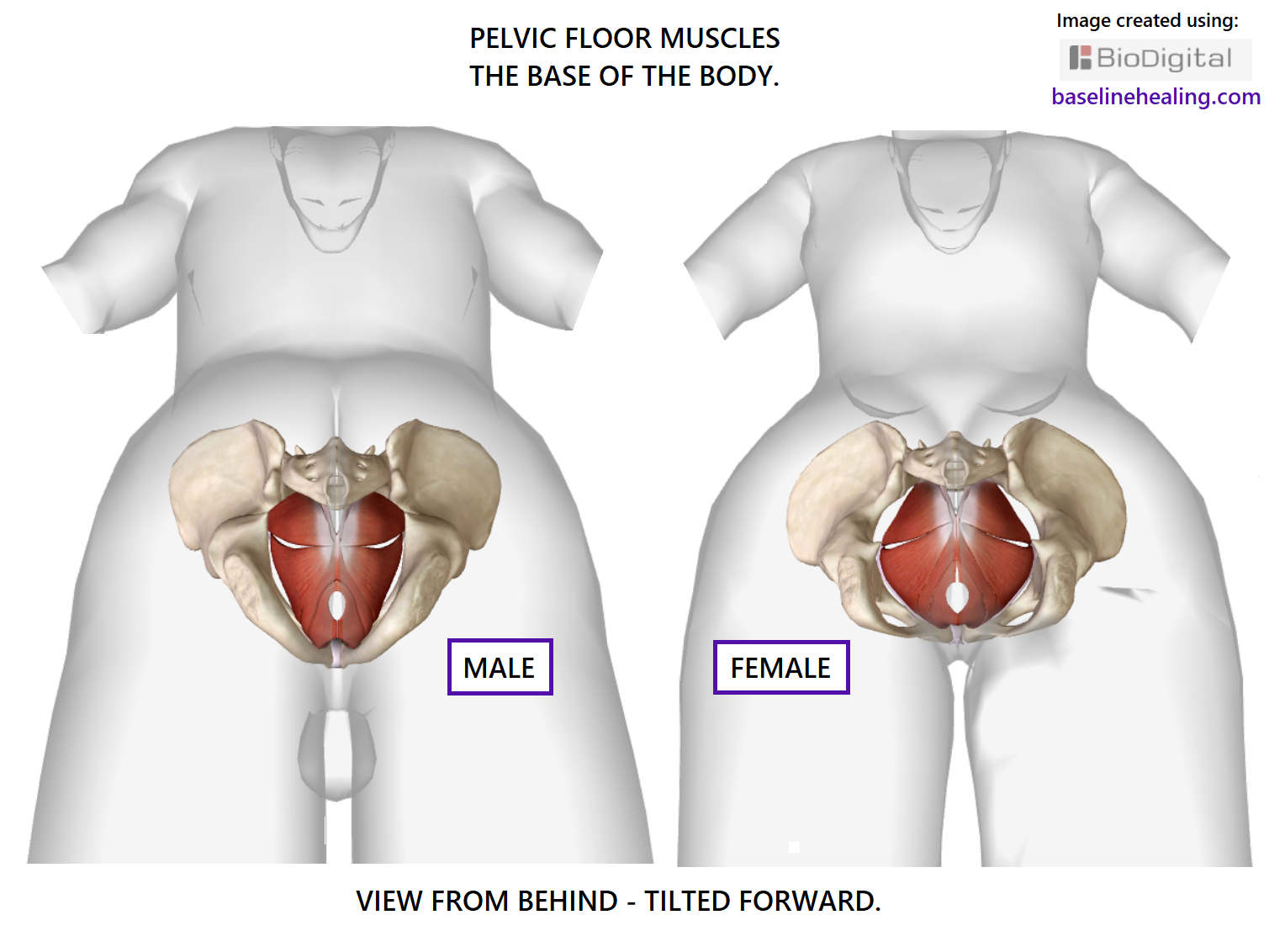
The pelvic floor - a crescent shape on the body's midline.
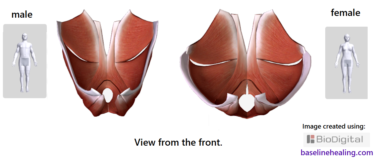
These muscles are closely associated with the anus and genitals.
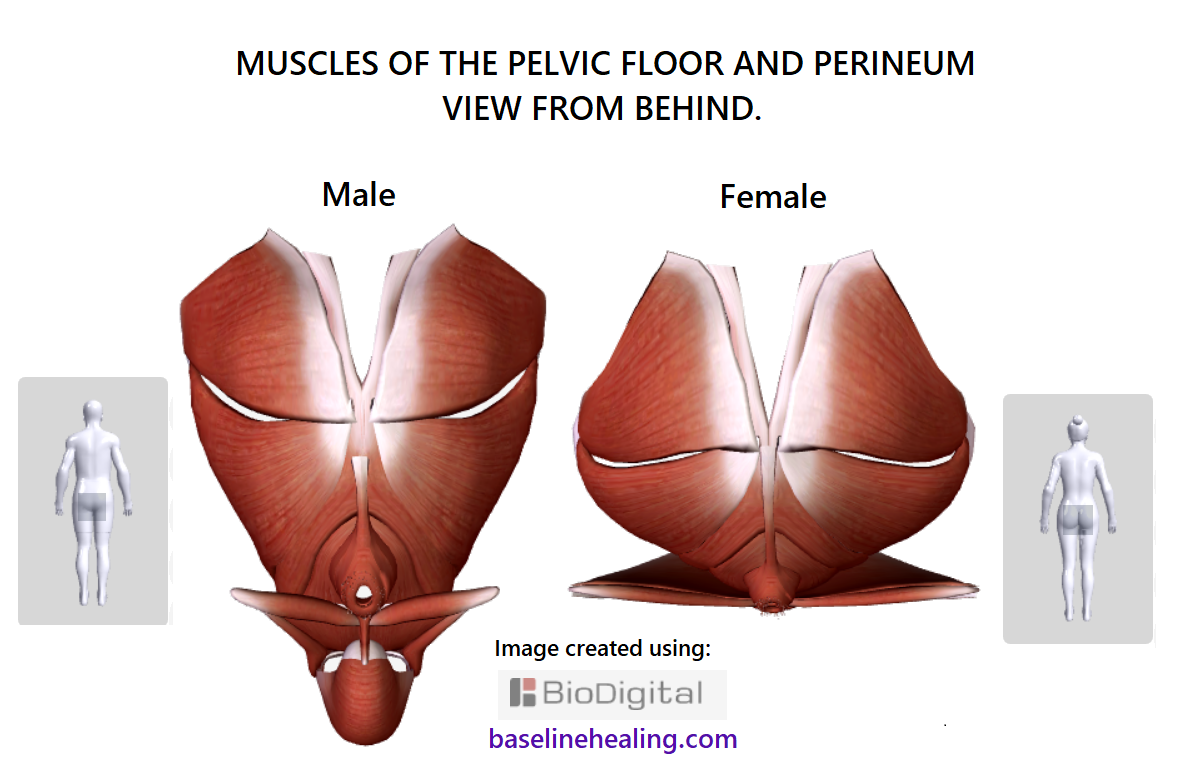
Kegel exercises are the most well-known introduction to strengthening the pelvic floor muscles. Picture these muscles contracting in your mind, feel for them working. Find a connection. Keep working at it. It will become easier the more you practice.
Aim for a balanced contraction left and right sides. Providing the sensory information for the 'base' point of our 'body map [LW · GW] in the mind' from where the rest of the body extends.
[The pelvic floor is generally accepted to correspond to the "root chakra [LW · GW]".]
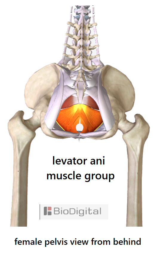
The pelvic floor muscles.
The base of the body.
4. Rectus abdominis. LINE
"The abs" = rectus abdominis muscles.
Strong and powerful, the muscles that allow the body to bend and flex in all directions when functioning at optimal.
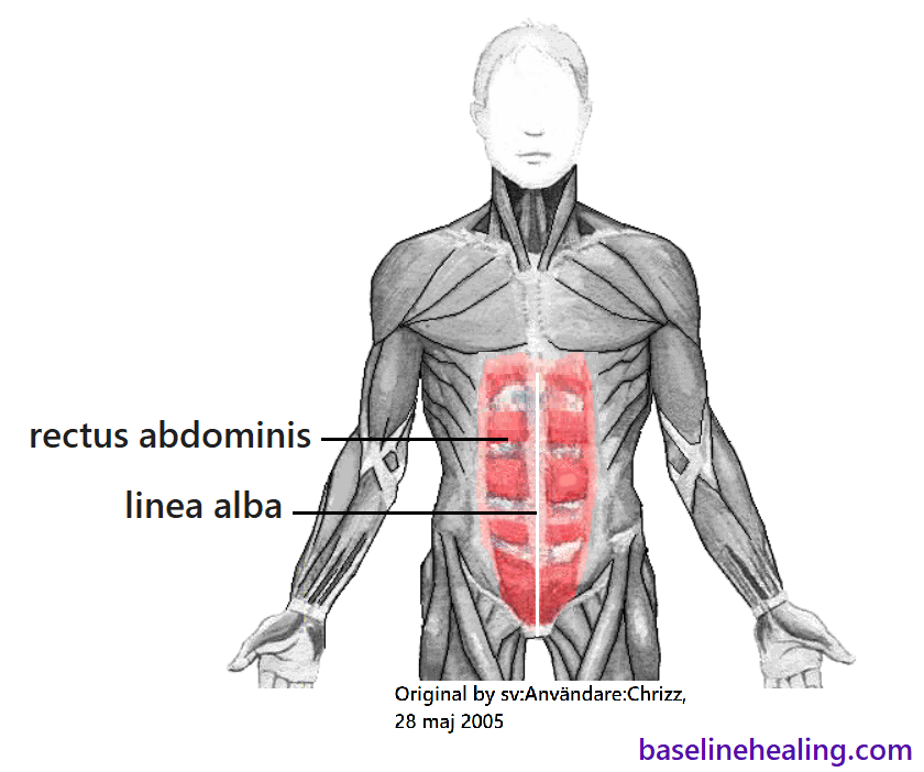
Think of these muscles as your central LINE, extending from a solid Base, that should be free to move and fully extendable.
The rectus abdominis muscles:
- From pelvis to chest up the front of your abdomen.
- Either side of the linea alba [LW · GW] - our primary guide for body alignment.
- Feel the relative positioning of 3 midline markers for body alignment [LW · GW] - the pubic symphysis, navel and xiphoid process of the sternum by working with your Base-Line muscles.

- The rectus abdominis consist of 2 parallel strips of panels of muscle within connective tissue. These panels create the "6 pack look" but the number of sections of muscle varies - 4, 6, 8, 10 packs can occur.

Feel your rectus abdominis muscles:

Place your hands over your rectus abdominis muscles, starting from the bone between your legs (pubic symphysis) then, as you breathe in, move your hands up thinking of activating and elongating - section by section - all the way up to your chest. (Put your hands over the front of your lower ribcage to appreciate how high up the chest the rectus abdominis attach).
Repeat the breathing and extension of your rectus abdominis, lengthen the muscles as much as you can and think about the linea alba straightening and aligning.
Think of the panels of muscle as a set of lights to be activated in sequence. Or whatever works for you ...
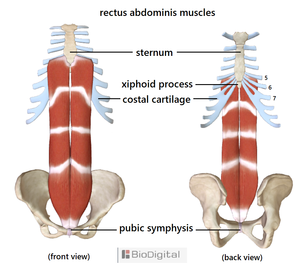
The rectus abdominis muscles - our core pillar of strength from where all movement should originate.
Breathe with your Base-Line [LW · GW].
Think stronger and longer with every breathe in.
5. Trapezius
A blanket of muscle that should be smooth and wrinkle-free, from mid-back to the back of the skull, extending out towards each shoulder.
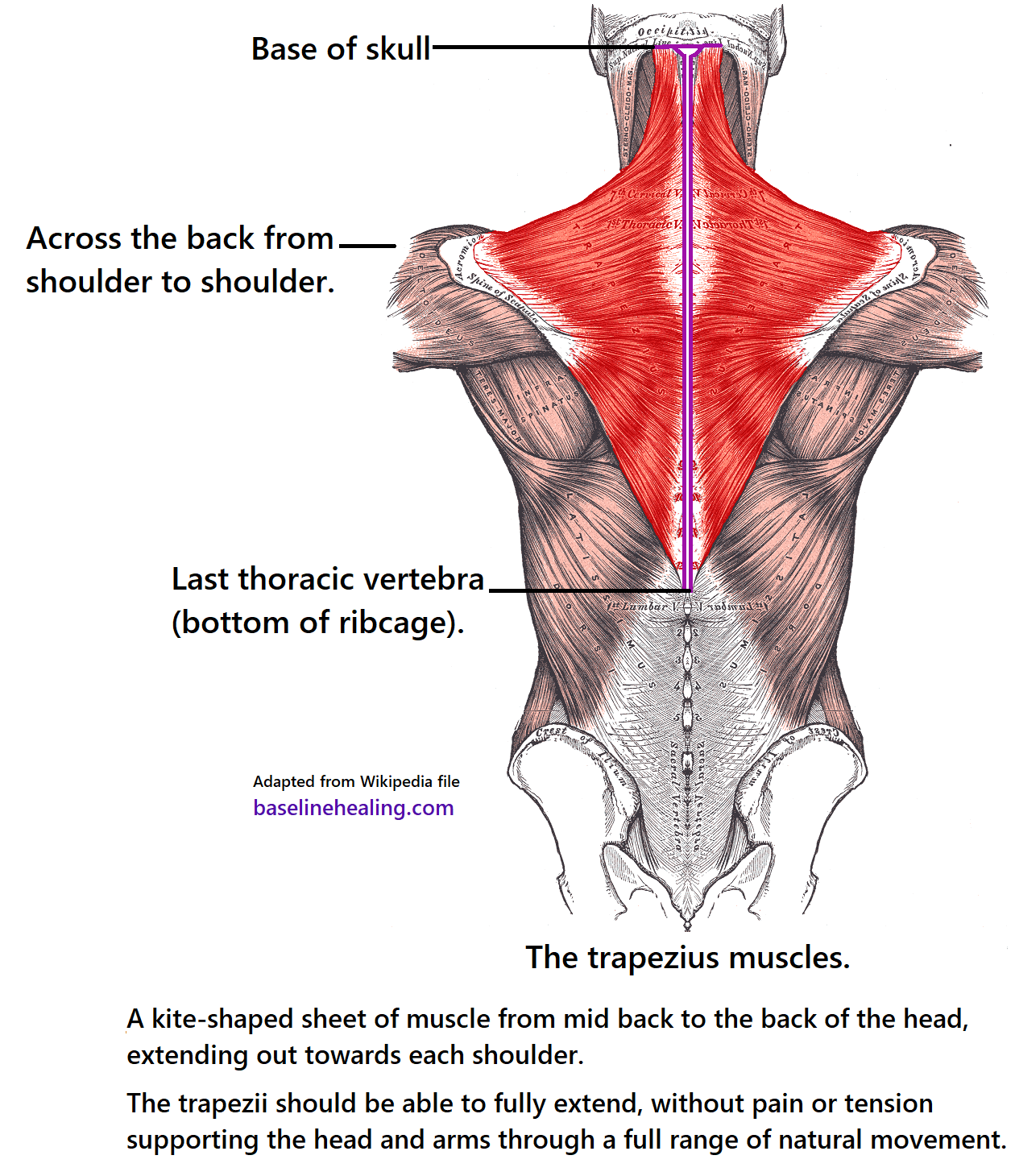
- Can you drop your head forward, extending from mid-back, without tension?
- Can you spread your arms wide, from midline to fingertips without restriction?
- Can you lift your arms up above your head feeling the trapezius muscles fully extend?

The trapezius muscles can be thought of as 6 sections (approximating - 2 triangles and a horizontal strip to the shoulders on each side as shown above).
- Mid-back, feel from bottom of the rib-cage extending up.
- Extending out to each shoulder.
- Feel for all the bony bits where the trapezius attaches near the shoulder.
- a 'pencil' like bone at the front (the collar bone/clavicle).
- lumps of bone at shoulder and a ridge of bone at the back. (parts of the shoulder blade/scapula).

- Sculpted over the front of the collar bone and up the sides of the neck.
- The trapezius muscles attach to the back of the skull. Feel for the ridge and midline bump at the back of the skull.
- The is a connective tissue 'ellipse' as the trapezius muscles meet in the upper back/between the shoulders region.
Movement of the upper body should begin from the lower trapezius.
Like wings extending from the middle of your back.
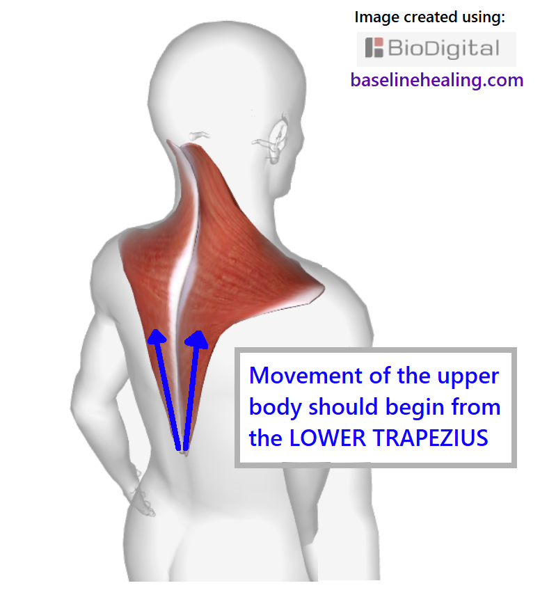
Think of lifting your shoulders from below, rather than pulling them up.

The trapezius muscles meet midline and merge with the nuchal/supraspinous ligaments [LW · GW] - our 'secondary guides for alignment'.
The trapezius muscles - guiding and supporting the head and arms through a full range of movement and aligning the upper body.
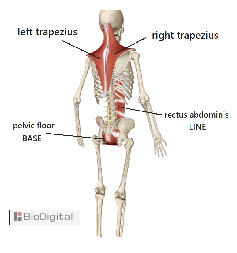
BODY ALIGNMENT with the main muscles of movement:
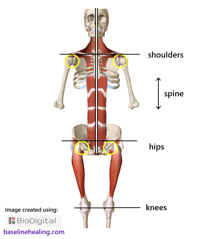

Find your 5 main muscles and work towards regaining a full range of natural movement, releasing the physical tensions on your body. The key to better health.

Time and Effort Required.
- Find these 5 muscles on your body.
- Be guided by your Base-Line and get moving. Use the roll-down [LW · GW] action.
- Feel for engagement and balance as you work with these muscles.
- Develop the connection between muscles and mind.
Link to 3D model on biodigital.com.
0 comments
Comments sorted by top scores.